Back to Back Blog

The Physical Therapies:- Osteopathy, Chiropractic and Physiotherapy ….The Differences…
Osteopathic therapy, physiotherapy, chiropractic care and massage share a common philosophy: The integrity of the spine is important in ensuring good health. In fact, this philosophy is shared by almost all traditional healing arts and is also found in many modern alternative treatments.
The Difference Between Osteos, Chiros, Physios and massage……
We are often asked how osteopathic therapy differs from physiotherapy, chiropractic care or massage.
Please note – this is general overview that we have found over the years and what many other patients have reported. All practitioners treat differently and it is important to find one that works for you! Being osteopaths – we are obviously Osteopathically biased!
Let’s start with the similarities:
Osteopathic therapy, physiotherapy, chiropractic care and massage share a common philosophy: The integrity of the spine is important in ensuring good health. In fact, this philosophy is shared by almost all traditional healing arts and is also found in many modern alternative treatments.
Now for the differences:
Generally people seek a therapist because of pain or impaired movement. Let’s look at how the same problem might be treated by the different types of therapists. Imagine you have a shoulder injury. You play some recreational golf and each year you get a twinge in your shoulder at the beginning of the season. You’d like to play golf pain-free and you’d like the pain dealt with once and for all.
You try physiotherapy . . .
Your treatment time will vary from 15 to 30 minutes.
The physiotherapist assesses your shoulder using standard orthopaedic tests and reaches the conclusion that there is some impingement of one of the rotator cuff muscles, which is a very common shoulder injury.
The therapist might choose to use some ultrasound on your shoulder.
You will get some specific exercises to increase strength to any weakened muscles of your shoulder.
The treatment may or may not include hands-on work. If it does, it will probably just be focused on your shoulder or upper ribs.
You are asked to come back twice a week for eight treatments.
You try chiropractic care . . .
Your treatment time will vary from 5 to 30 minutes for your first appointment, and last for about 5 minutes in subsequent sessions.
Like the physiotherapist, the chiropractor might assess your shoulder using some standard orthopaedic tests. The tests might also include an assessment of your spine, often using x-rays.
The chiropractor will be looking at the parts of your spine where the nerves to the shoulder come out, checking for what is called a subluxation. From the chiropractic perspective, the spine can become minutely out of alignment, and the resulting subluxations inhibit nerve flow, which can cause joints to become injured.
Treatment will probably involve manipulating your spine to free up the nerves so that they can control your shoulder better.
You are probably asked to come back two to three times a week for three weeks. You will then slowly decrease the frequency of your treatments until you are on some sort of monthly maintenance program to check for general subluxations.
You try massage . . .
Your treatment time is usually an hour.
The massage therapist will probably feel what muscles are tight and will just work on those muscles.
Generally you will lie down and the therapist will massage the tight or sore muscles to increase blood flow to the area to speed up the healing.
You will probably be encouraged to come back whenever you feel the need.
You try osteopathy at our clinic . . .
Your first treatment lasts one hour. Subsequent treatments last 30 minutes.
We assess your shoulder to find our which areas are damaged or strained.
We then assess your spine to see if the nerves to your shoulder are compromised. In this way we are similar to a chiropractor.
BUT we also look further afield because your shoulder never works in isolation:We assess other joints that work in conjunction with your shoulder, especially your wrists, elbows, neck and hips.We might even choose to look at movement patterns. For example, we’d ask you to demonstrate your golf swing technique to see if any restrictions in your hips or neck are placing too much work on your shoulder. We may even video this and slow it down.
The treatment is strictly hands-on.
The hands of an osteopathic therapist are more sensitive and knowledgeable than any type of machine. We don’t use ultrasound or any other machine to help us understand what’s going on.We find we get the best results by keeping the treatment to the work of our trained hands.We are trained to do manipulations (both gentle and firm), joint movements and massage techniques.We also know how to use our hands in a very subtle way to gently free restrictions around organs and other deep body structures to restore health to your whole body.It is key for us to grasp why your body is struggling and assessing and treating it in an joined-up way is important here. We may work with your hips and feet to enable you to achieve better range in your shoulder (as everything is connected….)
The course of treatment with osteopathic therapy
We might ask you back in one or two weeks.Dependent on what the diagnosis is or how severe the injury may be, on average, we will want to see you about four to six more times over a two to three month period to make sure the problem is resolved.That will also give us a chance to assess and help you with any other problems that we think will cause you trouble in the future.
Blog post by James Dodd

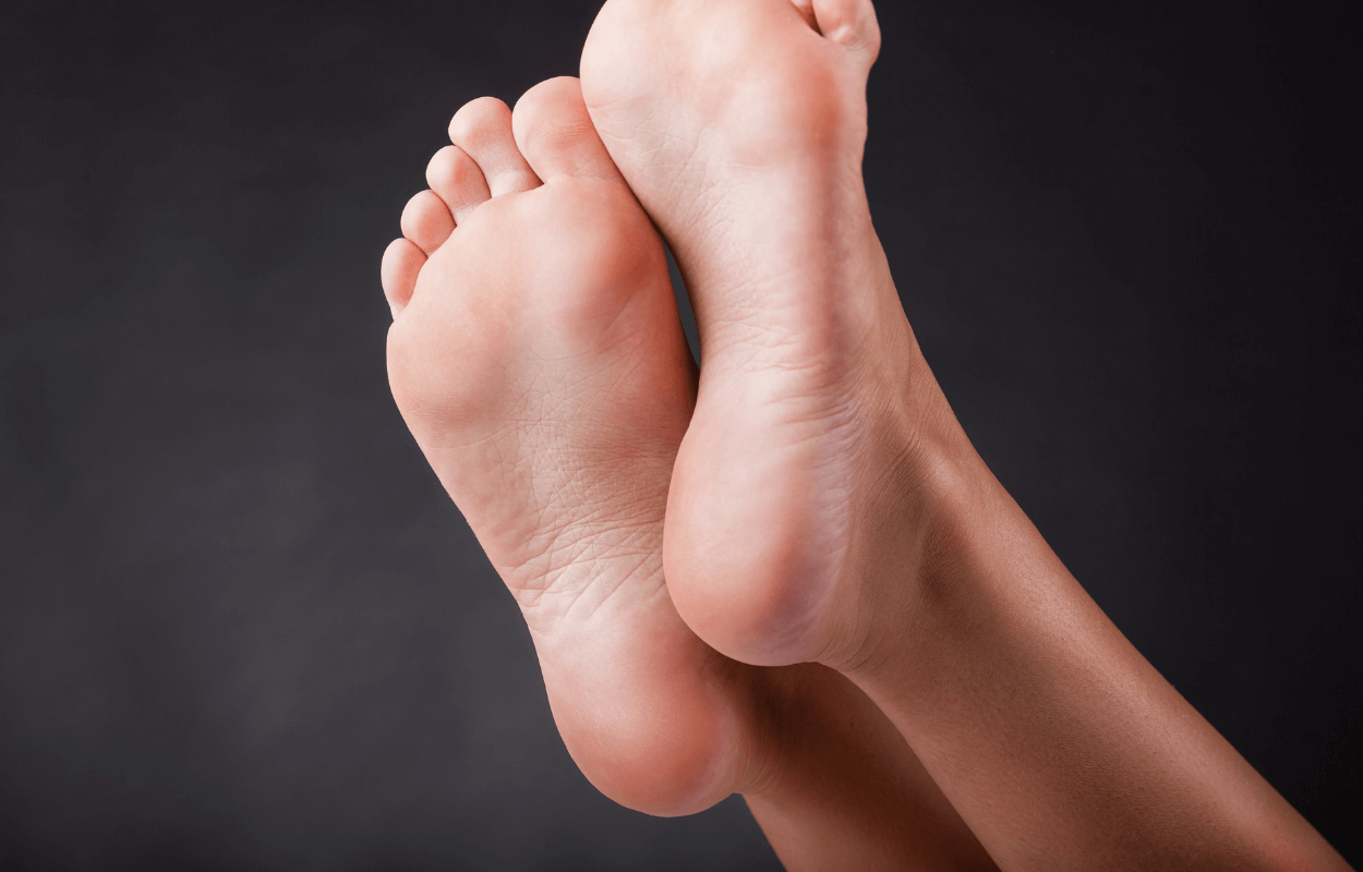
Sprained ankle on the tennis court? Think TWICE before grabbing the ICE
If you sprain your ankle on the tennis court, the first port of call is usually the clubhouse freezer. But is ice actually doing you more harm than good?

If you sprain your ankle on the tennis court, the first port of call is usually the clubhouse freezer. But is ice actually doing you more harm than good?
R.I.C.E. (Rest, Ice, Compression, Elevation) has been the most common initial treatment of acute injury over the last 30 years, as introduced by Dr. Gabe Mirkin in his 1978 publication The Sports Medicine Book. A recent study by the American Journal of Sports Medicine (June 2013), however, has made Dr. Mirkin swallow a slice of frozen humble pie. The study demonstrated no evidence that ice hastened recovery, leading Dr. Mirkin in his 2014 article “Why Ice Delays Recovery” to admit that he was wrong.
So, why is ice not always the best solution?
Firstly, it is worth noting that the inflammatory process is vital for the repair and remodelling of tissues. Common sense would suggest that inhibiting this process may not be the best idea. Ice acts to constrict blood vessels thereby reducing the amount of inflammatory cells deposited by your blood stream.
Although this may reduce pain and pressure on an injury, it also stops healing cells from entering injured tissue. Ice, as well as constricting blood vessels, also constricts the lymphatic system which is responsible for clearing out inflammatory debris. So, you can begin to get a picture of the effect ice has on an injury; less healing cells and a reduced ability to remove inflammatory waste – not ideal for recovery.
So, what should you do?
Here are Dr. Mirkin’s new set of tips for acute injury treatment:
1. Stop exercising immediately; you don’t want to cause further damage.
2. If the injury is very painful, then cold has been shown to reduce pain, in these circumstances you can grab a bag of peas from the freezer but use intermittently – 10 minutes on, 20 minutes off.
3. As soon as possible, get yourself assessed by a health professional to ensure no serious damage has been done.
4. After 48-72 hours the inflammatory process will usually have done its job, movement and the correct exercises then become the order of the day.
5. Joint pumping is a fantastic way of naturally assisting the lymphatic system to remove excess waste, while the correct movements will stimulate tissue repair.
If you are suffering from an injury and need to have it treated, just call the clinic to book an appointment with me.
Neil Sharland
Osteopath M.Ost

ACUPUNCTURE – THE ONLY RECOMMENDED PROPHYLACTIC TREATMENT FOR HEADACHE
Research has shown that a course of acupuncture can reduce symptoms of headaches by more than 50% and in some people acupuncture has been reported to eliminate their symptoms altogether!!

Suffering with a headache is unnecessary…
“Headache has been underestimated, under-recognised and under-treated throughout the world” WHO
Headache disorders are among the most common disorder of the nervous system. Nearly 50% of us have suffered from a headache in the last year and nearly 10% of those have reported migraine. Up to 4% of the worlds adult population suffer with headaches on 15 or more days a month.
Not only is headache painful, but it is also disabling.
Acupuncture for headaches…
Research has shown that a course of acupuncture can reduce symptoms of headaches by more than 50% and in some people acupuncture has been reported to eliminate their symptoms altogether!!
Following this research the National Institute for Clinical Excellence (NICE) passed a Guideline for Headaches, CG15 in September 2012 declaring a course of up to 10 sessions of acupuncture over 5-8 weeks as the ONLY recommended prophylactic treatment that isn’t drugs.
There are many types of headaches, some chronic and some episodic. Like with any other aches and pain the longevity of symptoms can act as a general guide of how many treatments you may need to help with your headache management. The more acute headache sufferer may only need 1-2 treatments, but for the more chronic and long-term sufferers, more treatments will be necessary. What is clear is that acupuncture is very likely to help with fighting your headache.
What to do next?
Start by keeping a headache diary today – record the frequency, duration and severity of the headache. Contact us if you would like a simple spreadsheet to follow.
If you are suffering with headaches, call the clinic to book an appointment in with me.
Anja Davidson
Osteopath M.Ost

Nutrition tips for marathon runners
Nutririon tips for marathon runners…

RUNNING THE MARATHON?
Here are some vital nutrition tips:
1. STAY HYDRATED
It’s really important to be hydrated for the marathon. The trick is in the preparation. You don’t want to be making lots of toilet trips during the race! Drink plenty the week before and in the morning 700ml will do, avoiding any for the hour before the race.
Make sure you have water throughout the run, but don’t feel the need to drink constantly. The most recent research suggests it’s best to drink when you are thirsty. This helps avoid over-hydrating and can also reduce gut discomfort and improve performance on the day. Coconut water is also a great natural alternative to the energy drinks in replacing lost hydration, sugars and electrolytes during the race.
2. FAT THEN CARB LOADING GIVES MORE ENERGY
Research has shown that a diet (short term) high in fat before you embark on the more traditional pre-race carb loading offers great benefits for increased energy. 10 days of fat loading are enough to increase the muscles fat burning capacity, while the three day carbohydrate load ensures muscles have plenty of glycogen available for energy.
In the fat loading days, start 2 weeks before the race and aim for 65% of your total calorie intake from foods containing healthy fats. These could be avocados, cheese, eggs, salmon, whole milk, Greek yoghurt, nuts, olives and olive oil. In the carb loading days, start 3 days before your race and aim to get 70% of your total calories from carbohydrates.
3. DRINK BEETROOT JUICE
Beetroot juice is packed with dietary nitrates, which dilate blood vessels, increasing blood flow to muscles during exercise. Studies have shown that drinking 500ml of beetroot juice 2-3 hours before running can enhance performance. Try it on a couple of training runs before your marathon day and see if the red juice helps.
REMEMBER YOUR NUTRITION IS FUEL
Getting it right can really make the difference not only in performance, but your enjoyment of the day. Good luck and fuel smart.
Written by Stephanie Gammell M.Ost FAFS
Functional Osteopath at Back to Back – The Earlsfield Osteopath

SHIN PAIN IN RUNNERS EXPLAINED
Lower leg pain can come on for unseasoned runners or those that change their training routine too quickly without laying suitable foundations. This could be switching to lots of hill running or adding in more speed work.
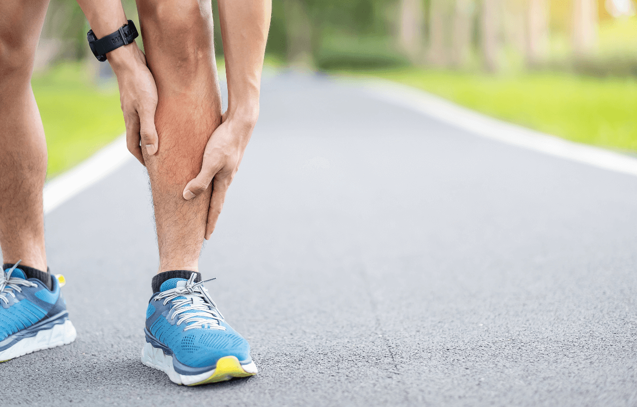
Lower limb injuries in runners are all too common and unfortunately hard to predict. With the ‘Virgin Money London Marathon’ not too far off, we wanted to share some Back to Back thoughts on shin pain.
Lower leg pain can come on for unseasoned runners or those that change their training routine too quickly without laying suitable foundations. This could be switching to lots of hill running or adding in more speed work.
One or more of THREE pathological processes are often involved in shin pain.
Shin Splints or Medial Tibial Stress Syndrome/Inflammatory shin pain
This is normally pain on the front or inside of your tibia/shin bone. It can wax and wane, but normally decreases as you warm up. The runner can often complete their training but it can recur after exercise and be painful the following morning. If left untreated, it can become worse.
It is generally agreed that if you have shin splints, you should stop running or alter your training depending on its severity. Reduction of the inflammatory response is key and it may be helped by rest, stretching, ice and soft tissue work.
Medical Acupuncture in the right places appears to be pretty effective. Off load your shins with alternative training methods or running in a pool. When you return to running, do it gently and follow the 10% rule. Don’t increase your speed or distance by more than 10% per week.
Bone Stress Response
Pain in the shinbone may be due to a stress response/stress fracture of your tibia. This without doubt is more serious than ’shin splints’ and needs to be ruled out if pain persists. This sort of pain can be increasing or pretty constant. It is often worse on impact or after use. There may be some night pain. Pain is normally more localised or acute than ‘shin splints’.
Compartment Syndrome
The muscles in your lower leg are separated into compartments. Causes are not fully known, but as your muscles swell during activity, they create increased pressure in these ‘closed compartments’. Signs and symptoms are directly related to use and intensity. It increases with exercise and decreases with rest. Soreness can be minimal and diffuse. There may be muscle weakness and sensory symptoms into the foot and toes.
Seeking help is important if you have pain, especially if it does not go away. Making sure you see an appropriate practitioner with suitable qualifications to enable a correct diagnosis or referral is important.

Cancaneal Apophysitis (Sever’s Disease)
Sever’s Disease in children; the cause and treatment.
This is painful inflammation of a child’s growth plate at the heel. Normally, this affects children between 8 – 14 years old as their calcaneus (heel bone) is still developing. It is also at a time when children often increase the amount of exercise they do. With increased and repetitive use, the achilles tendon ‘tractions’ on the growth plate at the heel and so causing pain and inflammation. Approximately 60% of Sever’s is bilateral.
Causes
It is essentially an overuse injury at the time of growth. Sports that ‘load’ the achilles tendon and heel such as running and jumping are normally the culprits. Often a bout of Sever’s can become aggravated at the start of a season after a ‘rest’ period or exercising on harder ground as it gets colder. Tightness in the calf can also lead to increased load onto the heel bone. It bad cases, it may take until the child stops growing before complete resolution. It is also really important to try to observe why there has been more load placed onto their heel… this may be from a stiff hip or other area. This is key to successful treatment.
Diagnosis
This needs to be based on a full and correct examination by your osteopath, doctor or other medical professional. X-ray or MRI may be used to confirm the diagnosis or monitor the progress, but often this is not necessary.
Treatment
Calcaneal apophysitis has no known long-term complications and is self-limiting; that is, it should go away when the two parts of bony growth eventually join together (occurring around 16 years of age).
It is important to limit (temporarily) excessive or rigorous activity in its painful stages. But it is also about management, as you can get times when it calms and at other times, it can then flare up again as they increase activity. Soft shoes and heel cups can make a difference and it is important to make sure the child has sound biomechanics (eg no excessive pronation or muscular imbalance). Regular and correct stretching of the tight muscles in the calf and thigh are essential. Ice can be of great help if used correctly. Anti inflammatory medication may be of use – but do check with your medical professional about this first.
Seeking help from your osteopath or good physical therapist can really help too. They will check for poor biomechanics and work and stretch the calf and thigh and manage this injury with some good strengthening exercises.
Return to sports or activity
The goal here is to get your child back to their desired sport or activity as soon as safely possible. It may be a gradual return to see if the condition regresses. If they return too early, it may lead to more chronic pain.
To return to sport your child should have no pain at rest and should be able to walk pain free. They should also be able to jog, sprint and hop pretty much symptom free too.
If after the pain resolves…. it is important that there is still a regime of regular stretching of their calves, thigh and leg muscles

Joint Replacements and osteopaths
Arthritis can affect people both physically and mentally and the pain it may cause can be extremely draining.

There are a number of patients who often present to our Osteopathy clinic for the treatment and management of arthritic conditions that may eventually require a joint replacement. Often these clients will ask the Osteopaths for their opinion on this and whether it is the right thing to do. This is often a difficult question to answer because every individual is different. Arthritis can affect people both physically and mentally and the pain it may cause can be extremely draining.
Other than advice from a health professional, it is also important to speak to others that have also had the same procedure. These are big operations and the ‘pros’ and ‘cons’ must be looked at. Most importantly, if you do decide to undergo an operation like this, you must be prepared to do the rehabilitation afterwards. This gives your body the very best chances of healing well and coping with your new joint.

Effective Spinal Manipulation….. what osteopaths do best!
Techniques between Osteopaths, Chiropractors and physiotherapists can differ hugely, but spinal manipulation is what we do best!
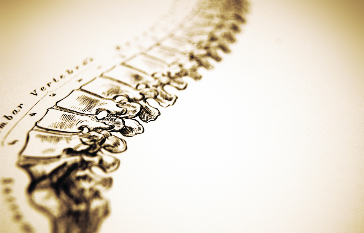
An interesting paper here on the effectiveness of spinal manipulation….
The results of this study confirm that lumbar mobilisation treatment has an immediate effect in relieving low back pain, however the specific technique used seems unimportant. More research is probably needed here to find about more specific techniques and how they work….. Techniques between Osteopaths, Chiropractors and physiotherapists can differ hugely and this needs to be reviewed too!

Biomechanics and REAL function
I have been an Osteopath since 1999 and part and parcel of being an osteopath is having a thorough understanding of anatomy, biomechanics as well as medicine and pathology.

I have been an Osteopath since 1999 and part and parcel of being an osteopath is having a thorough understanding of anatomy, biomechanics as well as medicine and pathology.
This year I had the privilege of doing a mentorship with the Gray Institute called GIFT. This was a 40 week course working with the world legends Gary Gray and Dave Tiberio. They created ‘Applied Functional Science’ or AFS. This is the real science behind movement and not what is says in the anatomy books! Gary Gray has been a US physical therapist for more than 35 years and has been working with Dave Tiberio since then. They look at REAL function and how everything changes when your foot hits the floor.
Assessment and rehabilitation is all done in 3 planes and mostly standing, using the full impact of gravity and ground reaction! This uses correct neurological pathways as your proprioception is ‘switched on’ and so you are able to assess what the body is able to ‘functionally’ achieve. This is very different from feeling and seeing what a back, hip or knee does while lying on a table.
Very few people REALLY look at the body in the way they (or I do now) do. Some will look at the tri-plane movement of the foot and sub-talar joint, but they don’t link this to the hip or scapular or cervical spine in gait, hitting a golf ball or bowling. Gary Gray and Dave Tiberio teach all this to the extent of how your psoas affects your scapular or foot! Quite brilliant!
GIFT has been inspirational and an amazing journey and it has set me alight! It has given me more passion to further the osteopathic and functional model. GIFT is a huge investment both financially and in your time. But I looked at it as just that…. an investment. An investment in me and an investment in my patients. So worth it!
If anyone is interested in doing something like this, look at their website and if you cannot manage something so big as GIFT, look at one of their Chain Reaction courses or their online content.

Femoral Anteversion and Retroversion
It is important that when testing the ROM available of the hip, it should be tested in normal function, upright (not just lying on a plinth) and with the hip in a flexed and extended position, since movement may be possible in one motion, but restricted into the other.

This one is often so overlooked and surgeons often suggest that there is no clinical consequence…..and so don’t worry!
For the office worker or the sedentary person that does not have large physical requirements of their body, it may just be the silent hip that may or may not give rise to any symptoms.
BUT for the person that does a very physical job or the person who partakes in sport or the hard training athlete…. the apparent symptoms may be very different!
Normal hip internal rotation should be about – 35 degrees
Normal hip external rotation should be about – 45 degrees
It is important that when testing the ROM available of the hip, it should be tested in normal function, upright (not just lying on a plinth) and with the hip in a flexed and extended position, since movement may be possible in one motion, but restricted into the other.
One possible cause of increased hip internal or external rotation may be femoral neck anteversion or retroversion.
The normal neck of the femur is angled at 15 degrees anterior to the long axis of the shaft of the femur and the femoral condyles. An increase to this anterior angulation results in greater internal rotation (anteversion) available at the hip. Often the patients are seen to be ‘toeing-in’ Conversely, a decreased anterior angulation (retroversion) results in a greater amount of external rotation. Patients that ‘toe-out’ may have a retroverted hip.
One thing we as clinicians need to be aware about when seeing patients is that either of these can create neck pain, lower back pain (or many others) as well as hip, knee and foot pain. Looking for this does not take long and can be part of your normal thorough examination.
Imagine the runner that toes-in due to a right anterverted hip. As he runs he needs internal rotation at his hip and by toeing-in, he uses much if this internal rotation up. This would tighten up the frontal plane of the hip and further load your knee as it tries to cope with the lack of available motion at the hip. Also, as the toes points inwards, your knee is not aligned in the sagittal plane. The creates excessive load at your knee too!
So be aware of this as a potential problem and check out some great and simple tests that may give you a hint as to whether this may be the root of your patients symptoms.
If you are concerned about your gait or hips, give Back to Back a call on 020 8605 2323 and one of us can have a look.

Adductor Function
They are the adductor brevis, longus, magnus oblique and magnus vertical. The brevis and longus attach onto the posterior medial part of the femur, not just on the medial part as most people talk about.

I just LOVE this muscle!
It does most things other than adduct your hip!! If you lie on the ground on your side and lift you lower leg… sure… your adductors adduct your hip…..BUT during function, it does a fabulous job at not adducting your hip.
They are the adductor brevis, longus, magnus oblique and magnus vertical. The brevis and longus attach onto the posterior medial part of the femur, not just on the medial part as most people talk about.
A great and massively overlooked thing about this group of muscles is that they work with their opposite adductors. The right and left adductors are turned on in gait (differently) but at the same time.
If the right leg is forward the right adductors are stretched in the sagittal plane and they slow hip flexion. They are lengthened in the transverse plane (TP) and they help to internally rotate the femur. They are also shortened in the frontal plane (FP) with hip adduction.
As we walk, and as the left leg swings forward, the right leg becomes the back leg and the right adductors are lengthened by hip extension (posterior medial attachment). They are then lengthened in the FP by the pelvis leaning towards to the left leg causing hip adduction. It is then shortened in the TP are they externally rotate the femur. The facilitates top-down external rotation of the tibia and calcaneal inversion…. and locks out the mid tarsals ready for push off!
As I have said – both sets of adductors work as a pair…. The right adductor works with the left adductor to slow the movement of the pelvis to the left and visa versa. If the adductors and tight in any plane, they will inhibit other planes.
But they can also be responsible for other dysfunction. If the adductors are short or not permitting good function, your pelvis will be unable to move correctly in 3 planes of motion and so your lower back, mid back or neck may take the hit instead. You might end up seeing someone for your back pain all because of you adductors. This is why it is SO important to not always treat the symptom, but to go to the cause!!
If you have just injured yourself (especially after the marathon) or need to be assessed for injury or need treating, do give us a call at Back to Back on
Hamstring function – This is not just a knee flexor!

Hamstring function – How it assists in knee extension
While on the subject of knee extension….
The hamstring can flex the knee against gravity when prone or standing with the foot in non-weightbearing….but during most function, the action of the HAMSTRING is so much more complex.
During swing phase, the hip is flexing and the knee is extending. Both of these motions lengthen the hamstrings so that it is switched on before heel strike. At heel strike, the forward motion of the trunk tries to flex the hip. This is decelerated by the hamstring. At the same time, the tibia is moving forward with knee flexion, which the hamstrings can slow down when the foot is on the ground.
As the heel bone everts, the lower leg internally rotates and because of its greats attachment onto the lower leg and the pelvis, the hamstrings can decelerate both internal rotation of the tibia and the hip. In the frontal plane, the hamstrings also decelerate abduction of the knee after the heel has everted.
The lengthening and eccentric loading of the hamstring at multiple joints in many planes make it very eccentric with the front leg in gait. But once the ‘unload’ has started, the hamstrings become very concentric in all planes.
All of this eccentric control (loading) allows it then to do the opposite (explode) once it has been stretched and proprioceptively ‘turned on’.
So in function and gait…. the HAMSTRING is used so much more than to flex your knee as the books suggest.

Hypermobility part 2 (March 2012)
Part 1 of this article looked at giving readers a better understanding of Hypermobility Syndrome (HMS) and the implications it may have on the musculoskeletal system. Having a greater understanding of the common problems associated with hypermobility syndrome, how it is diagnosed and its relationship to other connective tissues disorders provides us with a solid base by which we can then go about developing a corrective exercise program. Part 2 aims to provide a more extensive look at assessing the hypermobile patient and taking a region specific approach to training.

Hypermobility Syndrome – part 2
James Dodd
Part 1 of this article looked at giving readers a better understanding of Hypermobility Syndrome (HMS) and the implications it may have on the musculoskeletal system. Having a greater understanding of the common problems associated with hypermobility syndrome, how it is diagnosed and its relationship to other connective tissues disorders provides us with a solid base by which we can then go about developing a corrective exercise program. Part 2 aims to provide a more extensive look at assessing the hypermobile patient and taking a region specific approach to training. A detailed case history and screening process should alert you to any problematic areas or regions which may be predisposing, maintaining or aggravating your patient’s musculoskeletal problems. Static observation will help you to define any asymmetries in posture. Observation from all three views (frontal, lateral and posterior) will help you focus on areas of possible dysfunction. Functional testing in sagittal, frontal and transverse planes may highlight other areas of dysfunction
Muscle and Movement Testing
Carry out stretches and tests to identify weak, tight and shortened muscles. Take particular care in the hypermobile patient as range of movement may be much greater than expected. They are also good at ‘cheating’ to hide a dysfunctional area.
Assess functional activities such as gait, step up/down, lunge, squat, one leg stand and reach, anterior reach, posterior reach and push up for functional capability. Look for asymmetries, imbalances and glitches within the movement patterns to alert you to potential areas of dysfunction. Common causes of dysfunctional movements could be inhibited/over facilitated muscle activity in agonist/antagonist or synergistic muscles, tight and short or weak and lengthened muscles, hypo/hypermobile joints or body regions and balance, proprioception and coordination problems.
Always remember to seek advice from your patient’s general practitioner, osteopath or physiotherapist if you suspect any serious possible musculoskeletal problems or red flags.
A Regional Approach to Training the Hypermobile Patient
Common presenting features in the hypermobile patient include the following:
Shoulder, hip, neck, lower back, knee and foot pain
Poor body awareness, balance and proprioception
Flat, pronated feet
Hyperextended knees
Anterior pelvis
Clicking/popping joints
Hyperkyphotic or reduced movement in the thoracic spine. This could also be chronic muscle spasm
Tight and shortened hamstrings, gastrocnemius, iliotibial band, tensor fascia lata, pectoralis major/minor muscles
Weak anterior deep neck muscles, iliopsoas, core stabilizers and gluteals
Poor core stability
Upper rib breathing and poor diaphragmatic breathing
A good case history, observation and assessment should highlight any areas of dysfunction and will help you to focus on devising a program that will help restore optimal health and posture. Foam rollers, static stretching, active stretching and Muscle Energy Techniques (MET) are treatment strategies that can help stretch, activate and restore muscle balance. Remember to train the patient as a whole. The body works as one, and all systems are connected. Therefore, any one problem or area can predispose to a dysfunction elsewhere. The tissues are generally weaker and more vulnerable to injury than the non-hypermobile patient.
Hypermobility and the Lower Back
Poor overall functional stability is common among those individuals with lower back pain. In conjunction with poor stability, the hypermobile patient may also have poor kinesthesia and proprioception.
Focus training on:
Improving body awareness (anterior and posterior tilting of the pelvis)
Finding neutral spine
Teaching diaphragmatic breathing
Activating core muscles
Incorporating core muscle activation into functional activities
Balance, proprioception and coordination
Look for an ability to activate the core muscles:
Without co-contraction of more global muscles such as the rectus abdominis and external oblique muscles
Independently breathing
Diaphragmatic breathing? (often you will observe upper chest breathing)
Exercises that will be useful for the patient with core stability issues include:
Anterior and posterior tilting of the pelvis (standing, supine).
Core muscle activation (supine, prone, side-lying and four-point kneel).
Core muscle activation standing, seated.
Remember to isolate muscle activation and then integrate into functional activities.
Hypermobility and the Neck
Hypermobile patients may present with a history of neck complaints, ranging from the acute ‘wry’ neck to chronic neck pain. Individuals may present with an upper crossed syndrome pattern, which could be predisposed, maintained and aggravated by hypermobility, work and lifestyle postures such as sitting and slouching.
Look for:
Forward head posture (chin poking forwards)
Increased kyphosis (rounding) through the thoracic spine and Cervico-Thoracic C7/T1 region (base of the neck)
Reduced mobility through the thoracic and cervical spine
Tight upper trapezius, levator scapular, pectoral minor, sternocleidomastoid and scalene muscles
Weak rhomboids, lower trapezius and deep anterior neck flexors
Winging, elevated or protracted scapulars. Look for asymmetries in static
observation, movement and muscle weakness in shoulder stabilizers
Focus training on:
Lengthening short and tightened muscles
Strengthening weak muscles
Activating inhibited and de-facilitating over active muscles
Balance, proprioception and coordination
Exercises that will be useful for the patient with upper crossed syndrome and neck pain include:
Scapular retraction and setting
Ys and Ts exercise
Other useful exercises that aim to improve mobility of the neck, strengthen deep anterior neck flexors and increase body awareness include:
Chin tucks (similar to an emu neck movement)
Isometric neck flexion (using soft ball to push head against)
Isometric lateral neck flexion (using soft ball to push head against)
Pulling shoulder blades backwards and down, whilst lifting sternum to the roof
For more information on isometric neck exercises, see Paul Chek’s “How to Eat, Move and Be Healthy!”
Hypermobility and the Shoulder
The shoulder is the most unstable joint in the body, and often the hypermobile person will have a glenohumeral joint and/or scapulothoracic joint dysfunction. Problems may develop due to poor neuromuscular control, increased laxity of the capsule and rotator cuff weakness. Individuals may also present with subacromial impingement signs and symptoms. In some circumstances, your patient may require referral to an osteopath or physiotherapist to rehabilitate and teach correct scapulothoracic setting and glenohumeral positioning before commencing an exercise program. Focus training on:
Scapulothoracic setting
Rotator cuff strengthening
Stretching tight and shortened muscles i.e. pectoralis major and minor
Strengthening weak muscles i.e. serratus anterior, rhomboids, lower trapezius
Training functional exercises which address adjacent areas (cervical, thoracics, lumbars and hip/pelvis), muscular slings, synergistic and agonist/antagonist muscles
Hypermobility and the Hip
Like the shoulder, the hip is a ball and socket joint that has a large range of movement. It is essential that the neuromuscular, active and passive systems are working effectively to allow adequate movement and yet stabilize the region sufficiently. The hypermobile person is more likely to suffer from clicking/popping hip. The exact cause of this could vary from an intra to extra articular causes such as a tight iliopsoas to a labral tear or loose body. It may or may not be associated with pain. In those individuals presenting with extreme or constant pain, refer them off to the GP, sports medical doctor, osteopath or physiotherapist for further assessment. Typically, hypermobile patients will have poor control of their hip, pelvic and lumbar regions, and these often need to be addressed to correct any imbalances and prevent further problems developing.
Focus training on:
Lumbar (lower back) stability
Hip stability
Weak iliopsoas
Weak gluteus medius and maximus
Tight hamstrings, iliotibial band, tensor fascia lata
Balance, proprioception and coordination
Hypermobility and the Knee
Patellofemoral problems are common in hypermobile individuals. The cause of patellofemoral problems can be both structural and non-structural related. Common causes include a variable Q angle, genu varum, increased foot pronation and biomechanical and muscle imbalances.
Focus training on:
Tight iliotibial band, tensor fascia lata, hamstrings, gastrocnemius
Weak vastus medialis, gluteal muscles
Pronation and supination foot
Balance, coordination and proprioception
Exercises that will be useful for the client include:
Seated heel presses (helps to activate vastus medialis)
Foam rolling ITB and quadriceps muscles
Exercises that will be useful for the patient include:
Seated heel press (helps to activate vastus medialis)
Lunge – Forward
Lunge – Multiplanar
Squat – Against Wall with SB
Squat Touchdown – 1 Leg
Step Up to Balance – Frontal Plane
Hypermobility and the Foot
Pronated (flat) feet is common in the hypermobile patient. Over pronating can lead to problems with the subtalar joint, mid and forefoot, possibly causing plantar fasciitis and other problems up the biomechanical chain. Over pronation will cause internal rotation of the tibia and fibula, therefore potentially creating dysfunctions further up the chain at the knee, hip/pelvis and lower back. Individuals with flat feet may benefit from an exercise program or orthotics prescription.
Focus training on:
Tight plantar fascia, gastrocnemius and soleus muscles
Activation and control of tibialis anterior, peroneal muscles
Tight iliotibial band and weak gluteal muscles
Balance, coordination and proprioception
Exercises that will be useful for the patient include:
Active plantarflexion and dorsiflexion of the foot
Rolling feet inwards (pronation) and outwards (supination)
Single leg balance (stable to labile surface i.e. wobble board, bosu ball)
Swiss ball squat with ball between the knees finishing with standing on toes
Exercises that will be useful for the patient include:
Side to Side Hip Swing (Hip)
Balance Hold 1 Leg – Overhead Anterior Reach
Balance Hold 1 Leg – Overhead Posterior Reach
Hypermobility and Balance and Proprioception
Hypermobile individuals are likely to have poor balance, proprioception and kinesthesia. Exercises that challenge these components will translate over to improvement of general daily activities and reduce the likelihood of injury.
Exercises that will be useful for the patient include:
1 Leg Balance
1 Leg Balance Reach (anterior, posterior, lateral)
Balance Hold 1 Leg – Overhead Anterior Reach
Balance Hold 1 Leg – Overhead Lateral Reach
Step Up to Balance – Frontal Plane
Equipment such as wobble boards, Bosu Balls, cones, Airex Balance Pad, Swiss ball, Theraband, etc. can be added to further challenge balance and proprioception.
References:
Cook, G. Athletic Body in Balance, Human Kinetics, USA, 2003.
Gray, G. Functional Video Digest Series
Gray, G. Total Body Functional Profile, Wynn Marketing, 2001.
Janda, V. Muscle Function Testing, Butterworths, London, 1983.
Keer, R. & Grahame, R. Hypermobility Syndrome, Recognition and Management for Physiotherapists, Butterworth/Heinemann, 2003.
Kendall, F et al. Muscle Testing and Function with Posture and Pain, Lippincott Williams & Wilkins, 5th Edition, USA, 2005.
Murtagh, J. General Practice, McGraw Hill, 3rd Edition, Sydney, 2003.
Osar, E. Complete Hip & Lower Extremity Conditioning.
Osar, E. Complete Shoulder & Upper Extremity Conditioning.
Petty, N.J & Moore, A.P. Principles of Neuromusculoskeletal Treatment and Management, Churchill Livingstone, London, 2004.
Santana, J.C. Functional Training: Breaking the Bonds of Traditionalism
Wolf, C. Human Motion: A Pictorial Guide to Functional Integrated Movement Patterns. Human Motion Associates.
Resources:
www.ehlers-danlos.org <http://www.ehlers-danlos.org>
www.hypermobility.org <http://www.hypermobility.org>
www.marfan.org <http://www.marfan.org>
www.marfanssyndrome.net <http://www.marfanssyndrome.net>
www.medicinenet.com/hypermobility_syndrome <http://www.medicinenet.com/hypermobility_syndrome>
http://medlineplus.gov/
www.oif.org <http://www.oif.org> (Osteogenesis Imperfecta)
www.nlm.nih.gov/medlineplus/osteogenesisimperfecta.html <http://www.nlm.nih.gov/medlineplus/osteogenesisimperfecta.html>
Important Disclaimer:
No express or implied warranty (whether of merchantability, fitness for a particular purpose, or otherwise) or other guaranty is made as to the accuracy or completeness of any of the information or content contained in any of the pages in this web site or otherwise provided by personal training on the net. No responsibility is accepted and all responsibility is hereby disclaimed for any loss or damage suffered as a result of the use or misuse of any information or content or any reliance thereon. It is the responsibility of all users of this website to satisfy themselves as to the medical and physical condition of themselves and their clients in determining whether or not to use or adapt the information or content provided in each circumstance. Notwithstanding the medical or physical condition of each user, no responsibility or liability is accepted and all responsibility and liability is hereby disclaimed for any loss or damage suffered by any person as a result of the use or misuse of any of the information or content in this website, and any and all liability for incidental and consequential damages is hereby expressly excluded.

Hypermobility pt 1 (January 2012)
Part 1 of this article looked at giving readers a better understanding of Hypermobility Syndrome (HMS) and the implications it may have on the musculoskeletal system. Having a greater understanding of the common problems associated with hypermobility syndrome, how it is diagnosed and its relationship to other connective tissues disorders provides us with a solid base by which we can then go about developing a corrective exercise program. Part 2 aims to provide a more extensive look at assessing the hypermobile patient and taking a region specific approach to training.

Hypermobility Syndrome – part 1
James Dodd
There is Hypermobility and Hypermobility Syndrome (HMS). Hypermobility syndrome is the hypermobile patient with symptoms. Not all hypermobile patients are, or ever become symptomatic. HMS is a condition many individuals often experience their entire lives without ever having it diagnosed and managed correctly. Hypermobility has certain implications for the joint itself, as well as the surrounding ligaments, bones, joint capsules, muscles and tendons. It is important for a hypermobile patient’s well-being that they have effective neuromuscular control and active and passive systems that can support the joints in the body. Hypermobile joints by definition are those “displaying a range of movement that is considered excessive, taking into consideration the age, gender and ethnic background of the individual.” Hypermobility Syndrome, also known as Joint Hypermobility Syndrome or Benign Hypermobility Syndrome, is defined as ‘generalized joint laxity with associated musculoskeletal complaints in the absence of any systemic disease’. HMS is an inherited form of a connective tissue disorder. Those with Hypermobility Syndrome are believed to experience pain as a result of joint microtrauma, which has been caused by overuse and/or misuse of the tissues in which there is an inherent weakness in the collagen.
It’s signs and symptoms are so often missed and so it goes undiagnosed and so is mismanaged. This means the suffering patient can go on suffering unnecessarily.
Common Features of Hypermobility
•Females are often more mobile than males.
•Mobility varies amongst ethnicity (Asian populations seem to be more affected than European populations).
•Mobility decreases with age.
•Hypermobility can be inherited or acquired i.e. repeated subluxation of the shoulder joint.
•Hypermobility Syndrome is typically determined by the genetic make up of the individual, in particular the genes causing hypermobility are those that are responsible for the production of collagen, elastin, fibrillin and tenascin.
•Certain sports and activities (i.e., gymnastics, ballet dancing, swimming) have shown to have a greater number of individuals with hypermobility.
•Musculoskeletal regions commonly affected include the achilles tendon, patellofemoral joint, rotator cuff, carpal tunnel, cervical and lumbar spine (non specific lower back pain and medial and lateral epicondyles.
•Hypermobile individuals may suffer from clicking, spondylolithesis and pars fractures.
•Hypermobility is a risk factor for the development of early Osteoarthritis.
•Fibromyalgia is commonly diagnosed in individuals with Hypermobility Syndrome
Diagnosis
The Beigton Score System (below) is a 9-point scale originally introduced to recognize hypermobile patients within the population. Primary health care practitioners now use it as a common screening process to help determine whether an individual has hypermobility.
•Passively dorsiflex 5th metacarpophalangeal joint to > 90 degrees (2 points)
•Passively take thumb to forearm (2 points)
•Passive hyperextend the elbow to > 10 degrees (2 points)
•Passively hyperextend the knee to > 10 degrees (2 points)
•Actively place hands flat on floor without bending the knees (1 point)
There can be a total score of 9. The revised diagnostic criteria for hypermobility requires you to get a Beigton score of 4/9 and have arthralgia (joint pain) for longer than three months in more than four or more joints.
There is also the Brighton Criteria that aids in the diagnosis of Hypermobility Syndrome. These include:
Dislocation and/or subluxation in more than one joint and on one or more occasions.
Abnormal skin: striae, thin skin, increased stretch of skin.
Eye signs: drooping eyelid or myopia
Varicose veins, hernia, or uterine/rectal problems
Soft tissue rheumatism (i.e., epicondylitis, tenosynovitis, synovitis)
Hypermobility and Connective Tissue Disorders
Marfan’s Syndrome, Ehlers-Danlos Syndrome and Osteogenesis Imperfecta are each genetic disorders in which the connective tissue of the individual is affected. Each disorder is different in its epidemiology, incidence and clinical presentation. While it is beyond the realm of this article to discuss each of these conditions in detail, if you have a patient with one of these disorders, you should conduct further reading in order to gain a better understanding of the disorder, its implications and how you can alter your training program to better suit your patient.
Rehabilitation and Exercise Prescription
Management of the patient will largely depend upon his/her age, current physical condition, hypermobility state and personal goals. The primary aim of dealing with any hypermobile patient should be to reduce disability and increase function. This should incorporate a holistic approach which addresses not only the fitness components but also addresses dietary, work and other lifestyle components.
Hypermobility and Acute or Chronic Injury
The acute hypermobile client should be managed like any other acute musculoskeletal injury. During the acute stages, you should be aiming to reduce swelling, inflammation, pain and restrict any aggravating or maintaining factors. Always refer your patient to a medical practitioner or hospital if you suspect anything serious. In the sub-acute or chronic hypermobile patient, the aim should be to restore normal function and reduce pain to a comfortable level. This could range from prescribing an exercise program to referring your patient for specific treatment from a GP, Physiotherapist, Osteopath or Podiatrist. It is always best to seek treatment from a practitioner that that works with and understands the implications of HMS. Failing to correctly manage with your client’s sub-acute/chronic injury could result in the development of compensatory patterns, which in turn could lead to problems developing elsewhere.
Program Design
Training the hypermobile patient requires special attention to certain training principles. You need to consider all joint, muscle and ligament actions when training, making sure that you train throughout the entire range of movement and training the muscles for isometric, isotonic, concentric and eccentric actions.
Stability of a joint is determined by the integrity of the musculoskeletal system (muscle, tendon, capsule, ligament and articular surfaces) and the neural control system (motor and sensory). It is important to address these aspects when devising a program in order to develop optimum function.
The key fitness components that need addressing in the hypermobile client include:
Proprioception
Balance
Coordination
Kinesthesia
Core stability
Endurance
Strength
Controlled flexibility
Cardiovascular fitness
Each of these fitness components requires extra attention in clients with Hypermobility Syndrome. However, each program will differ considerably depending upon each patient (i.e., client age, current physical state, hypermobility and goals). Programs could range from being as simple as improving one’s functional state (i.e., daily living activities such as walking, squatting, sitting up and down) to training elite athletes. Essentially, you will need to assess each case individually and design a program specific to the client’s needs.
Your program should try to:
Train function. Include functional activities such as push/pull, step up/down, squat, lunge and rotation exercises in your program.
Strengthen muscles equally and ensure there is a balance among agonist, antagonist and synergistic muscles.
Train movements rather than muscles, when possible.
Progress from slow to fast, stable to unstable, simple to complex, non weight bearing to weight bearing, closed chain to open chain, one plane to multi plane.
In Part 2 of this article, I will be taking a more in depth look at the individual assessment of the hypermobile patient and how we can deal with the common musculoskeletal issues one may have. The article has been divided into bodily regions, outlining what we should be looking for in the assessment, how this interacts with other regions of the body and how we can go about correcting these problems.
References:
Cook, G. Athletic Body in Balance, Human Kinetics, USA, 2003.
Gray, G. Functional Video Digest Series <http:> </http:>
Gray, G. Total Body Functional Profile, Wynn Marketing, 2001.
Janda, V. Muscle Function Testing, Butterworths, London, 1983.
Keer, R. & Grahame, R. Hypermobility Syndrome, Recognition and Management for Physiotherapists, Butterworth/Heinemann, 2003.
Kendall, F et al. Muscle Testing and Function with Posture and Pain, Lippincott Williams & Wilkins, 5th Edition, USA, 2005.
Murtagh, J. General Practice, McGraw Hill, 3rd Edition, Sydney, 2003.
Osar, E. Complete Hip & Lower Extremity Conditioning.
Osar, E. Complete Shoulder & Upper Extremity Conditioning.
Petty, N.J & Moore, A.P. Principles of Neuromusculoskeletal Treatment and Management, Churchill Livingstone, London, 2004.
Santana, J.C. Functional Training: Breaking the Bonds of Traditionalism <http:> .</http:>
Wolf, C. Human Motion: A Pictorial Guide to Functional Integrated Movement Patterns. Human Motion Associates.
Resources:
www.ehlers-danlos.org <http:> </http:>
www.hypermobility.org <http:> </http:>
www.marfan.org <http:> </http:>
www.marfanssyndrome.net <http:> </http:>
www.medicinenet.com/hypermobility_syndrome <http:> </http:>
http://medlineplus.gov/
www.oif.org <http:> (Osteogenesis Imperfecta)</http:>
www.nlm.nih.gov/medlineplus/osteogenesisimperfecta.html <http:></http:>
Important Disclaimer:
No express or implied warranty (whether of merchantability, fitness for a particular purpose, or otherwise) or other guaranty is made as to the accuracy or completeness of any of the information or content contained in any of the pages in this web site or otherwise provided by personal training on the net. No responsibility is accepted and all responsibility is hereby disclaimed for any loss or damage suffered as a result of the use or misuse of any information or content or any reliance thereon. It is the responsibility of all users of this website to satisfy themselves as to the medical and physical condition of themselves and their clients in determining whether or not to use or adapt the information or content provided in each circumstance. Notwithstanding the medical or physical condition of each user, no responsibility or liability is accepted and all responsibility and liability is hereby disclaimed for any loss or damage suffered by any person as a result of the use or misuse of any of the information or content in this website, and any and all liability for incidental and consequential damages is hereby expressly excluded.
Could your groin injury be coming from your hip?

Groin pain can be a difficult problem for patients as well as their clinicians. Part of the problem is that the location of the pain is often a poor indicator of the where the pathology actually lies. Additionally, when the pain becomes chronic, multiple pathologies can be generated, adding a further layer of complexity.
It is very important to make a clear diagnosis, and we should seek to look beyond merely labelling the problem as a ‘groin sprain’. There many causes of pain in the groin, but approximately 50% of groin pain can be attributed to pain generated by the hip joint; a surprise, perhaps, for younger patients. As in any medical condition, the patient’s history will give us many clues. It is extremely important to rule out sinister ‘red flags’, such as night pain, severe pain on loading the leg, weight loss or systemic symptoms, and we need to be mindful of conditions which may occur in certain age groups, such as slipped epiphysis in teenagers.
Common causes of groin pain besides the hip, include those generated by the lumbar spine, pubic overload (osteitis pubis), iliopsoas and adductor tendon pathologies and stress responses in the femoral neck in runners. Abdominal wall hernias may cause pain which is a little higher in the groin, and less commonly, younger patients can experience the rectus femoris pulling away from its attachment at the anterior inferior iliac spine. Testicular tumours and avascular necrosis can present insidiously and we need to be on the lookout for them.
A big proportion of patients who present with groin pain as a result of hip pathology, have an underling condition known as ‘femoral acetabular impingement syndrome’, or FAI. This is essentially a problem resulting from a tear in the acetabular labrum, usually caused by repetitive trauma due to a ‘bump’ or ‘CAM’ on the head neck junction of the femur, which may be genetic.
This can cause groin pain which is worse with exercise, sitting or standing, and the pain can be brought on by putting the patient in the ‘impingement position’ of hip flexion + internal rotation + adduction. In the long term, we believe that the tear in labrum causes changes in the acetabular articular cartilage next to it, and over many years, this may lead to osteoarthritis in the hip.
FAI can affect people of all ages, and is often missed in 30-40 year olds. Taking a careful history, and carrying out a thorough examination can help identify the likely cause. Imaging, such as MRI arthrogram of the hip, can help confirm the underlying diagnosis (as X-Ray cannot rule out FAI), but it should be remembered that imaging needs to be interpreted in light of the history and examination findings. FAI may require treatment with hip arthroscopy surgery, but in some cases injection therapy and robust physiotherapy or osteopathy may be enough to get a person back to full activity.
Sports Physicians and Osteopaths are ideally placed to identify the underlying cause of unexplained groin pain, and are skilled in directing the rehabilitation necessary to resolve the problems.
If you have any problems at all and would like in to see James Dodd or one of the team at Back to Back, please call 020 8605 2323.